
Case Study: 5-Month-Old Kitten Diagnosed with Wet Feline Infectious Peritonitis
Today, I want to share a very interesting case: a 5-month-old kitten diagnosed with wet feline infectious peritonitis (FIP). Before discussing our patient in detail, I’ll give a brief overview of the cause of this feared disease.
Feline infectious peritonitis (FIP) is a serious condition in cats caused by a virus known as feline coronavirus (FCoV). There are two types of this virus: a more common and generally harmless type that affects the intestines, and another mutated type that can cause FIP. The disease develops when the intestinal coronavirus mutates within the cat’s body, allowing this mutated form to infect immune cells, which triggers a severe inflammatory response.
FIP manifests mainly in two forms:
- Wet or exudative form: Characterized by fluid accumulation in cavities like the abdomen or chest, causing symptoms such as abdominal swelling, difficulty breathing, and persistent fever.
- Dry or non-exudative form: In this form, there is no fluid accumulation, but granulomas (masses of inflammatory cells) form in various organs, affecting their function. Symptoms are more varied and include fever, weight loss, jaundice, and neurological signs if the brain is affected.
FIP is particularly dangerous due to the complexity of its diagnosis and treatment, and the disease is often fatal if not detected early. Although some promising treatments have been developed in recent years, prevention remains essential. This involves reducing stress and maintaining good hygiene practices in environments where multiple cats live, to minimize the spread of feline coronavirus.
Now that you have a clearer idea about this disease, let’s discuss our patient. A 5-month-old Scottish Fold came to the clinic showing symptoms of anorexia, lethargy, and dyspnea. This indoor cat was up-to-date on vaccinations and had been acquired from a breeder. After taking a history, we proceeded with the physical examination:
- Mental status: alert.
- Temperature: 39.2°C.
- Mucous membranes: pink, with capillary refill time under two seconds.
- Thoracic auscultation: no significant findings.
- Good hydration status; body condition score: 3/5.
Next, we started with complementary tests:
- Radiography: We performed two thoracic views, one ventrodorsal and one laterolateral, where we detected pleural effusion.
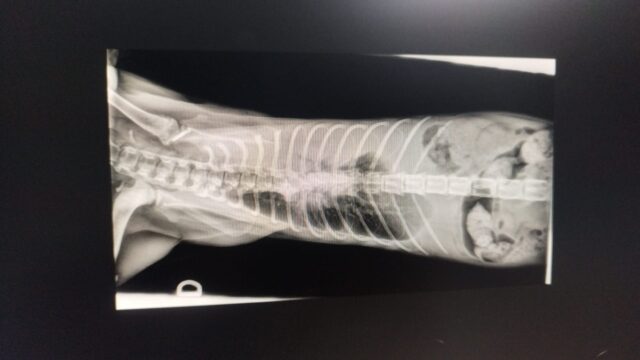
VD thoracic radiography 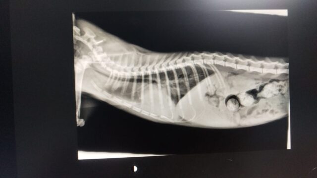
LL thoracic radiography - Thoracic ultrasound
: To obtain a sample of the pleural effusion.
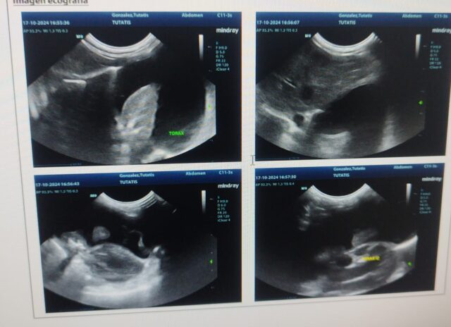
Thoracic ultrasound showing pleural effusion. - Laboratory tests
: A blood count showed moderate leukocytosis with neutrophilia and monocytosis. The biochemistry only revealed mild stress hyperglycemia.
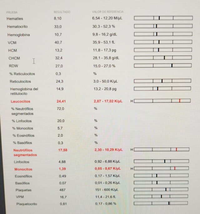
Our patient’s blood count showing leukocytosis with neutrophilia and monocytosis.
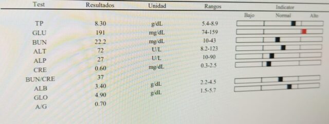
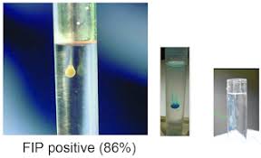
With these tests completed, we began to suspect an infectious cause. Our main suspects were FIP and feline leukemia, so we proceeded with a rapid FIV/FELV test, which was negative. After ruling out FELV, we performed the Rivalta Test on the pleural effusion sample, and it was positive.
As mentioned in the summary, diagnosing FIP can be very complex. When FIP is suspected, the Rivalta Test is crucial, as it is a simple, quick, and economical test that helps differentiate transudates from exudates (FIP, lymphoma, septic exudate). A positive reaction indicates a high protein content and the presence of inflammatory mediators and fibrinogen in the fluid.
The albumin/globulin ratio in the biochemistry can also be helpful, although it is not definitive. A ratio above 0.8 suggests FIP is unlikely, a ratio between 0.8 and 0.3 indicates a possible FIP, and a ratio below 0.3 indicates a high likelihood of FIP. Our patient’s ratio was 0.7.
I usually perform a cytology of the effusion sample, as its composition can also assist in the diagnosis. In FIP cases, we expect a high protein content (>3 g/dl), low cellularity (with only some degenerated neutrophils and macrophages), and sterile fluid, without bacteria.
With all these tests, we have a strong suspicion of FIP; however, we need something more conclusive. We sent a sample of the pleural effusion to the lab for a FIP PCR test. The value of PCR for FIP in pleural effusions is that a positive result has high diagnostic value, especially in cats with compatible clinical signs like persistent fever, fluid accumulation, and weight loss. However, a negative result does not rule out FIP, as the virus might be in tissues rather than the fluid. False positives can also occur but are rare when PCR is performed on effusions and combined with adequate clinical analysis.
In our patient, PCR returned positive for the S1060A mutation. The M1058L and S1060A mutations are the most studied as they are linked to changes in the spike protein, crucial for the virus’s ability to infect macrophages and spread systemically. However, the development of FIP is a multifactorial process that can involve multiple mutations throughout the viral genome, not just these two specific ones. Other identified mutations include those in the spike protein gene and in other viral genes, such as 3c and ORF1a.
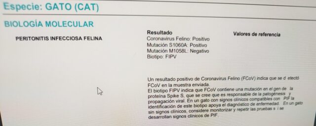
Once we had the definitive diagnosis, we could begin treatment. Until recently, there was no treatment for FIP, and it was considered a fatal disease. However, we now have an effective tool that has saved the lives of many FIP-positive cats: GS-441524. This antiviral, derived from the nucleoside adenosine, works by inhibiting viral RNA replication, preventing the virus from spreading throughout the body and helping reduce FIP symptoms.
Treatment usually lasts between 8 and 12 weeks, although it may extend depending on the cat’s severity and individual response, with survival rates of 80-90% in those treated early. While generally safe, side effects can include injection site pain and, rarely, hepatic or renal toxicity.
Currently, our patient is progressing favorably, although the treatment has not yet been completed, so we will need to wait a little longer to assess his evolution.
In another article, I will go into more detail about this promising new treatment.