
Leishmaniasis is a parasitic disease of great relevance in veterinary medicine, especially in endemic areas such as the Mediterranean. This pathology primarily affects dogs, although cats can also be affected to a lesser extent. Below, we will address its pathogenesis, symptoms, diagnosis, treatment, and preventive measures.
Pathogenesis
Leishmaniasis is caused by protozoa of the genus Leishmania, with Leishmania infantum being the most common species in Europe. The parasite is transmitted through the bite of sandflies (mosquitoes of the genus Phlebotomus), which act as vectors. During feeding, the sandfly inoculates infective promastigotes into the host. These promastigotes transform into amastigotes within macrophages in a period of 24 to 48 hours.
In the vector mosquito, promastigotes require 7 to 10 days from ingestion to reach the infective stage in their mouthparts, at which point the sandfly can transmit the infection to another host.
Once a dog is infected, the incubation period varies considerably, ranging from 3 months to several years before clinical signs develop. However, during this time, the animal can become a source of infection for sandflies.
The course of the disease depends on the host’s immune response. A strong cellular response (Th1) can control the infection, while an exacerbated humoral response (Th2) favors disease progression, manifesting with clinical symptoms ranging from skin lesions to severe kidney failure.
Symptoms
The symptoms of leishmaniasis may vary depending on the stage of the disease and the organ affected. The most common symptoms include:
- Skin signs: Exfoliative dermatitis, alopecia, hair loss around the eyes (“goggles” appearance), skin ulcers, and subcutaneous nodules.
- Systemic signs: Progressive weight loss, lethargy, intermittent fever, and generalized lymphadenomegaly.
- Renal signs: Chronic kidney failure manifested by polyuria, polydipsia, vomiting, and anorexia.
- Ocular signs: Uveitis, conjunctivitis, and keratitis.
- Other signs: Epistaxis, lameness, diarrhea, and muscle atrophy.
Diagnosis
The diagnosis of leishmaniasis requires an integrated approach, combining clinical history, clinical signs, and laboratory tests:
- Clinical examination: Weight loss, lymphadenomegaly, exfoliative dermatitis, ulcerative lesions, epistaxis, lameness, and signs of kidney failure.
- Hematology and biochemistry: Anemia, hyperproteinemia with hypergammaglobulinemia, and hypoalbuminemia. Advanced cases may show renal impairment.
- Serology:
- Indirect Immunofluorescence (IFI): Commonly used to detect antibodies.
- ELISA: Offers high sensitivity and specificity.
- PCR: Detects parasite DNA, especially useful in early stages or asymptomatic animals.
- Cytology or biopsy: Allows observation of amastigotes in macrophages from lymph node aspirates, bone marrow, or skin lesions.
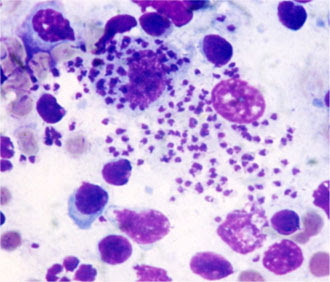 Amastigotes in macrophages.
Treatment
The treatment of leishmaniasis does not eliminate the parasite but controls symptoms and reduces the parasite load. It includes:
- Pentavalent antimonials: Such as meglumine antimonate (10-20 mg/kg SC, once a day for 4 weeks).
- Allopurinol: (10 mg/kg every 12 hours) inhibits parasite replication.
- Miltefosine: (2 mg/kg orally, once a day for 28 days) is often combined with allopurinol.
- Supportive treatment: Management of kidney failure, control of secondary infections, and nutritional supplementation.
Periodic follow-ups with serological and hematological tests are crucial to evaluate the treatment response.
Monitoring the Disease
Once leishmaniasis is diagnosed, regular follow-up is essential to assess disease progression and treatment efficacy. Recommended tests include:
- Complete blood count: To monitor anemia and detect potential secondary infections.
- Blood biochemistry: To evaluate kidney function (urea and creatinine), liver function, total protein levels, and the albumin/globulin ratio.
- Protein electrophoresis: To identify alterations in protein fractions, especially hypergammaglobulinemia.
- Quantitative PCR: To measure the parasite load in blood or tissues.
- Serology (IFI or ELISA): To evaluate antibody levels and their evolution.
- Urinalysis: Includes the calculation of the protein/creatinine ratio (UPC) to detect early kidney disease.
The monitoring protocol depends on the animal’s clinical status, but controls are generally recommended every 3-6 months.
Prevention
Since leishmaniasis has no definitive cure, prevention is key:
- Vector control: Use collars impregnated with deltamethrin or fluralaner and repellent pipettes.
- Vaccination: Available vaccines, such as CaniLeish® or Letifend, reduce the risk of developing clinical disease.
- Immunomodulators: Leishguard® stimulates the cellular immune response.
- Avoiding exposure: Keep animals indoors during peak sandfly activity hours (dusk and night) and use mosquito nets impregnated with insecticides.
Conclusion
Leishmaniasis is a complex disease that requires a comprehensive approach for its management. Combining preventive measures, early diagnosis, and appropriate treatment can significantly improve the quality of life of affected animals. As veterinarians, our role is to inform pet owners and work together to reduce the impact of this disease.